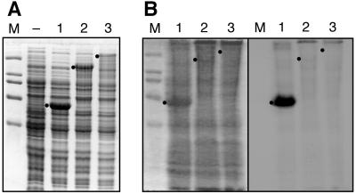
Figure 5.
Covalent incorporation of l-[3H]Leu into CepA proteins present in E. coli cell lysates. (A) Coomassie-stained gel showing expression of CepA1-575-His6 (lane 1); CepA1-1596-His6 (lane 2); and CepA1-3158-His6 (lane 3). (B Left) Coomassie-stained gel of cell lysates after treatment with Sfp, CoA, MgCl2, ATP, and l-[3H]Leu; and (Right) an autoradiogram of the same gel. l-Leu incorporation is detected for CepA1-575-His6-containing lysate (lane 1) but not for the larger proteins CepA1-1596-His6 (lane 2) and full-length CepA1-3158-His6 (lane 3). The expected or observed positions of the three proteins on the gels are indicated. Reactions contained (in a total volume of 120 μl) 50 μl lysate, 65 nM Sfp, 0.1 mM CoA, 10 mM ATP, 0.2 mM l-[3H]Leu (1.5 Ci/mmol), 50 mM sodium phosphate, 125 mM NaCl, 5 mM MgCl2, 10% glycerol at pH 7.5.

