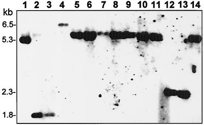
FIG. 2.
In vivo rearrangements of M. agalactiae avg-B2-bearing genomic fragments in successive isolates from a chronically infected ewe. HindIII-digested genomic DNAs from 13 clinical isolates (lanes 2 to 14) and from the M. agalactiae PG2 type strain (lane 1) were subjected to Southern blot hybridization with an avg-B2-specific oligonucleotide probe (pex-1). The isolates were obtained from a single naturally infected animal, designated animal #627, over a period of 7 months, and M. agalactiae was consistently isolated at each sampling time (at 2- to 4-week intervals) (10). The positions of molecular size markers are indicated on the left.

