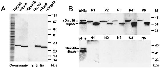
FIG. 2.
Western blot analysis of recombinant HpaA and Omp18 expressed in HK293 cells. (A) The purified recombinant antigens and lysate from untransfected HK293 cells were separated on a 4 to 12% Tricine-SDS-polyacrylamide gel and detected either by Coomassie blue staining (left lanes) or with a murine antibody against the His tag (αHis, 1:5,000) (right lanes). The secondary antibody was peroxidase-labeled anti-mouse IgG (1:20,000). The immunoblots were developed with a chemiluminescence system. The molecular masses are indicated on the right. (B) The purified recombinant antigens were separated on 10% tricine-SDS-polyacrylamide gels, transferred to nitrocellulose, and probed with five H. pylori-positive sera (P1 to P5, 1:500) or five sera from H. pylori-negative individuals (N1 to N5, 1:500). As controls, both recombinant proteins were detected with a murine antibody against the His tag (αHis, 1:5,000). The secondary antibody was peroxidase-labeled anti-human/anti-mouse IgG (1:20,000). The immunoblots were developed with a chemiluminescence system. The molecular masses are indicated on the right.

