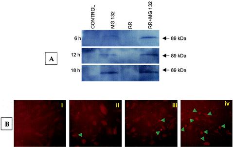
FIG. 2.
Cleavage of PARP during R. rickettsii infection of EC. (A) Western blot analysis for detection of cleaved PARP at different times after infection with R. rickettsii in the presence and absence of the NF-κB inhibitor MG132. (B) Immunofluorescent staining of cleaved PARP at 12 h in (i) uninfected EC, (ii) MG132-treated cells, (iii) cells infected with R. rickettsii, and (iv) R. rickettsii-infected cells in the presence of MG132. Arrowheads point to cells positive for PARP cleavage and morphological changes characteristic of apoptotic death.

