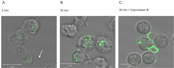
Figure 4.

Localisation of CFSE-stained Bb-HP incubated with human neutrophils. Confocal microscopy was used to demonstrate intracellular localisation of CFSE-stained Bb-HP. Neutrophils and CFSE-stained Bb-Hp were incubated for 5 min (A) or 30 min (B) before the reaction was stopped by adding ice-cold PBS and samples were fixed. The white arrow on panel (A) indicates a borrelia bacterium attached to neutrophil surface. In panel (C), neutrophils were pre-treated for 10 min with 5 μg/ml Cytochalasin B before addition of CFSE-stained Bb-HP for 30 min. Images shown are "stack images" taken at a defined cross section of the neutrophil, as close to the centre of the cell as possible. The white bar represents 10 μm. Images are representative of two to three samples carried out with neutrophils from two to three different donors.
