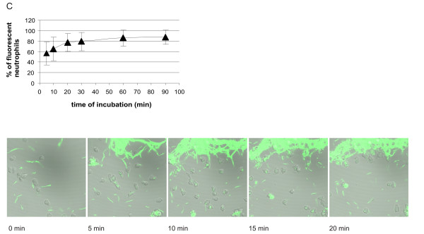
Figure 7.

Kinetics of association of CFSE-stained Bb-HP, Ba or Bg with human neutrophils. Neutrophils and CFSE-stained Bb-Hp (Fig. 5), Ba (Fig. 6) or Bg (Fig. 7) were incubated for 5, 10, 20, 30, 60 or 90 min before the reaction was stopped by adding ice-cold PBS and samples were fixed. Flow cytometry was performed using "cell settings" showing only fluorescence associated with neutrophils. Values are expressed as mean ± standard deviation and are obtained from seven to nine different experiments carried out with neutrophils isolated from six to eight different donors. Confocal microscopy was performed using live neutrophils and CFSE-stained Bb-HP (Fig. 5), Ba (Fig. 6) or Bg (Fig. 7). Scanning was performed at 37°C and images were stored at intervals of 2.5 seconds. Pictures show images taken at 0 (immediately after addition of bacteria into the culture dish mounted in the microscope), 5, 10, 15, and 20 min after addition of fluorescent bacteria. The tendency of Bg to form aggregates can be seen in the pictures. Images are representative of three to four "live-sessions" carried out with neutrophils from two to three different donors.
