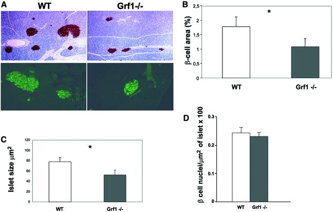
Fig. 6. Morphological analysis of pancreatic β-cells. (A) Representative insulin immunostaining of pancreatic sections from wild-type and knockout male mice. Upper panels: low magnification immunohistochemical analysis with anti-insulin antibodies. Lower panels: higher magnification of immunofluorescence studies. (B) The percentage of total pancreatic area occupied by β-cells was calculated using insulin-stained pancreas sections. For this analysis, three sections from each animal were immunostained and data were collected from eight independent fields of each section using Openlabs software. Results represent examination of sections from four wild-type and four knockout animals (16 weeks of age; P = 0.06). (C) Average islet size was assessed using pancreatic sections double-stained with glucagon and insulin. Measurements were performed using Openlabs software. Results represent examination of sections from four wild-type and four knockout animals (16 weeks of age; P = 0.03). (D) To approximate the density of β-cells/islet, paraffin-embedded pancreas sections were immunostained with insulin. To reveal the nuclei, sections were also exposed to DAPI. The area represented by insulin-containing cells was tabulated as described above, and nuclei were counted manually from these cells stained for insulin.

An official website of the United States government
Here's how you know
Official websites use .gov
A
.gov website belongs to an official
government organization in the United States.
Secure .gov websites use HTTPS
A lock (
) or https:// means you've safely
connected to the .gov website. Share sensitive
information only on official, secure websites.
