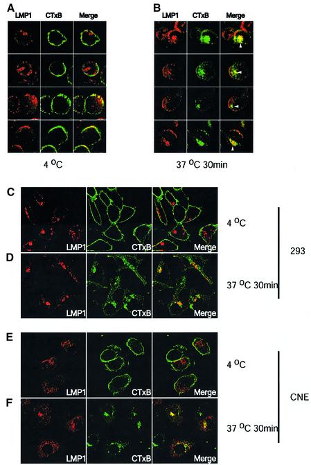
Fig. 1. LMP1 co-localizes with internalized cholera toxin. 721 cells, 293 or CNE cells transiently expressing LMP1 were incubated with 2.5 µg/ml FITC-conjugated cholera toxin (green) at 4°C for 30 min. Cells were washed twice with cold medium and either fixed, or shifted to 37°C for 30 min then fixed. The fixed cells were then stained for LMP1 (red). Yellow indicates co-localization of LMP1 and CTxB. At 4°C, most of LMP1 did not co-localize with CTxB in 721 cells (A, the first and second rows). In ∼16% of 721 cells, LMP localized to the cap-like structures which co-localized with surface-bound CTxB (A, third and fourth rows, see text). LMP1 also co-localized with internalized CTxB in 721 cells (B). White arrowheads in (B) indicate co-localization of LMP1 and CTxB at the perinuclear structures after CTxB had moved intracellularly on incubating live cells at 37°C for 30 min. In 293 and CNE cells, co-localization of LMP1 and surface-bound CTxB was rarely detected (C and E). LMP1 at the perinuclear and vesicular-like structures did co-localize with internalized CTxB (D and F).

An official website of the United States government
Here's how you know
Official websites use .gov
A
.gov website belongs to an official
government organization in the United States.
Secure .gov websites use HTTPS
A lock (
) or https:// means you've safely
connected to the .gov website. Share sensitive
information only on official, secure websites.
