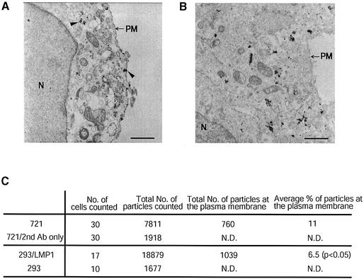
Fig. 3. Electron microscopy confirms that the majority of LMP1 does not localize at the plasma membrane. LMP1’s subcellular localization was examined in 721 (A) and 293 (B) cells by immunogold electron microscopy as described in Materials and methods. Arrowheads in (A) indicate examples of staining of LMP1 at the plasma membrane and at intracellular compartments. N, nuclear; PM, plasma membrane; scale bar = 1 µm. The fraction of LMP1 at the cell surface was quantified by counting the number of particles from randomly chosen cells (C). The difference between 721 and 293 cells was significant (Wilcoxon one-sided test, P < 0.05). ND, not determined. The silver enhancement of the fine gold particles leads to their dimensions varying. Each deposition was counted once independently of its size.

An official website of the United States government
Here's how you know
Official websites use .gov
A
.gov website belongs to an official
government organization in the United States.
Secure .gov websites use HTTPS
A lock (
) or https:// means you've safely
connected to the .gov website. Share sensitive
information only on official, secure websites.
