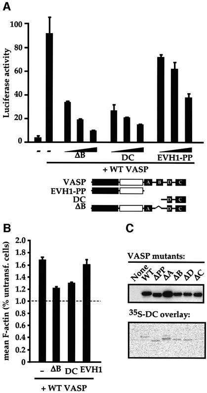
Fig. 3. The inactive VASP mutants ΔB and DC are interfering mutants. (A) Interference with VASP-induced SRF activation. NIH 3T3 cells transfected with the SRF reporter expressed intact VASP (0.15 µg) or the indicated VASP mutants (0.1, 0.3 and 0.9 µg). The structure of the mutants is shown below. (B) Interference with VASP-induced F-actin accumulation. Cells expressing intact VASP (0.75 µg) and the indicated VASP mutants (4.5 µg) were analysed for mean cellular F-actin content relative to that of untransfected cells in the same population using FACS. (C) The VASP EVH2 block C is sufficient to bind intact VASP. Lysates from cells expressing the indicated VASP mutants were separated by SDS–PAGE (12.5% gel), transferred to a membrane, and probed either with anti-Flag antibodies (upper panel) or with 35S-labelled VASP DC (residues 278–380), produced by in vitro translation (lower panel).

An official website of the United States government
Here's how you know
Official websites use .gov
A
.gov website belongs to an official
government organization in the United States.
Secure .gov websites use HTTPS
A lock (
) or https:// means you've safely
connected to the .gov website. Share sensitive
information only on official, secure websites.
