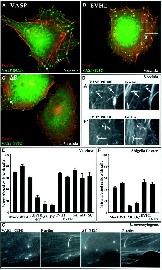
Fig. 4. Interfering VASP mutants block Vaccinia and Shigella F-actin tail formation. Adherent HeLa cells expressing VASP (0.3 µg) or its derivatives (0.6 µg) were maintained in 10% FCS. Sixteen hours later, the cells were infected for 8 h with Vaccinia (A–D, F) or for 4 h with L.monocytogenes (E) or S.flexneri (F), and processed for immunofluorescence. Cells were stained for F-actin using Alexa 568-phalloidin (red) and for the VASP epitope tag using 9E10 antibody (green). (A–D) VASPΔB blocks Vaccinia actin tail formation. Merged images are shown. Arrows indicate instances of VASP localization to focal adhesions. DAPI staining indicated the presence of viral particles at the tail ends (not shown). Quantitation is shown in (F). (A) Wild-type VASP; similar results were obtained upon infection of cells expressing GFP. (B) VASP EVH2. (C) VASPΔB blocks tail formation. (D) VASP and F-actin localization in the tails of cells expressing intact VASP (A′) or VASP EVH2 (B′). (E and F) Data summaries. The proportion of transfected Vaccinia-infected cells with any viral tails in a given field is shown. Data are represented as means ± SEM (n = 3). (E) Vaccinia data. In control infected cells expressing GFP, cells exhibiting tails generally contained 30–60 virus particles with tails. Expression of the interfering mutants reduced the proportion of cells displaying tails, and decreased the number of virus particles with tails to five to 10 per cell. (F) Shigella flexneri data. In control infected cells expressing GFP alone, those cells displaying tails contained only two to eight bacteria with tails. (G) VASPΔB expression allows the formation of L.monocytogenes actin tails. Separate images of VASP and F-actin are shown for infected cells expressing intact VASP (left) or VASPΔB (right). VASP derivatives did not affect the number of bacteria per infected cell. In two independent experiments, 58% and 55% (intact VASP) and 54% and 52% (VASPΔB) of infected cells displayed tails.

An official website of the United States government
Here's how you know
Official websites use .gov
A
.gov website belongs to an official
government organization in the United States.
Secure .gov websites use HTTPS
A lock (
) or https:// means you've safely
connected to the .gov website. Share sensitive
information only on official, secure websites.
