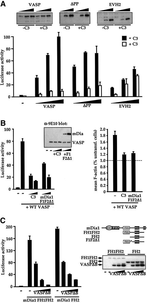
Fig. 6. VASP functions in the Rho–mDia–actin pathway to SRF activation. (A) Functional Rho is required for efficient VASP-induced SRF activation. Cells transfected with SRF reporter expressed intact VASP (0.02, 0.05, 0.15 µg) or VASP mutants (ΔPP: 0.02, 0.05, 0.15 µg; EVH2: 0.05, 0.2, 0.6 µg), together with C3 transferase (0.1 µg) as indicated. (Inset) Immunoblot analysis of VASP protein levels. (B) Functional Rho and mDia1 are required for VASP-induced SRF activation and F-actin accumulation. Left: SRF activation. Cells transfected with SRF reporter and expressing C3 transferase (0.1 µg) or interfering mDia1 mutant F1F2Δ1 (Copeland and Treisman, 2002) (0.9 µg) were processed as in (A). Inset: VASP immunoblot of cell lysates. Right: F-actin accumulation. Cells expressed intact VASP (0.75 µg) with either C3 transferase (0.5 µg) or mDia1 mutant F1F2Δ1 (4.5 µg). Mean cellular F-actin content of the transfected cells was determined relative to that of untransfected cells in the same population using FACS. (C) Functional VASP is required for efficient mDia1- induced SRF activation. Left: cells transfected with SRF reporter expressed activated mDia1 mutants FH1FH2 (0.03 µg) or FH2 (0.3 µg) (Copeland and Treisman, 2002), and VASPΔB (0.1, 0.3 and 0.9 µg). Right: mDia derivatives. Ellipse, Rho-binding domain; D, DAD domain. Immunoblot for mDia1 indicates that VASPΔB expression does not affect mDia1 protein levels.

An official website of the United States government
Here's how you know
Official websites use .gov
A
.gov website belongs to an official
government organization in the United States.
Secure .gov websites use HTTPS
A lock (
) or https:// means you've safely
connected to the .gov website. Share sensitive
information only on official, secure websites.
