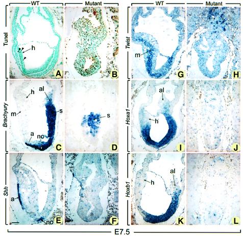
Fig. 4. TUNEL and in situ hybridization analyses of E7.5 normal and mutant embryos. Sections of E7.5 normal embryos (A, C, E, G, I and K) and their presumptive mutant littermates (B, D, F, H, J and L) were subjected to TUNEL reaction (A and B) and were hybridized with Brachyury (C and D), Shh (E and F), Twist (G and H), Hoxa1 (I and J), and Hoxb1 (K and L) antisense probes. In (A) and (B), brown-stained nuclei indicate end incorporation in DNA (arrowheads). Abbreviations: a, axial mesendoderm; al, allantois; h, head folds; m, mesoderm; no, node; s, primitive streak. Scale bar: 60 µm.

An official website of the United States government
Here's how you know
Official websites use .gov
A
.gov website belongs to an official
government organization in the United States.
Secure .gov websites use HTTPS
A lock (
) or https:// means you've safely
connected to the .gov website. Share sensitive
information only on official, secure websites.
