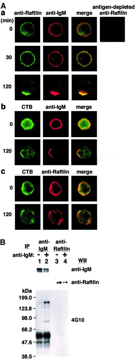
Fig. 4. Localization of Raftlin in Daudi cells. (A) Co-localization of BCR and Raftlin in Daudi cells. Cells were stimulated with anti-human IgM Ab for indicated periods and stained with a combination of anti-human Raftlin Ab and anti-human IgM Ab (a), or CTB and anti-human IgM Ab (b), or CTB and anti-human Raftlin Ab (c). Antiserum passed through a recombinant Raftlin protein column (antigen-depleted anti-Raftlin) was used as a negative control. (B) Immunoprecipitation of BCR and Raftlin using Daudi cells. Cells (6 × 10⁷) stimulated with or without anti-human IgM (20 µg/ml) for 5 min at 37°C were lysed with digitonin-lysis buffer and immunoprecipitated with anti-human IgM Ab and protein-G–Sepharose 4FF (Amersham Bioscience) or anti-human Raftlin IgG-immobilized Sepharose. Immunoprecipitants were divided into three aliquots and blotted with anti-human IgM, anti-human Raftlin and anti-phosphotyrosine Abs (4G10).

An official website of the United States government
Here's how you know
Official websites use .gov
A
.gov website belongs to an official
government organization in the United States.
Secure .gov websites use HTTPS
A lock (
) or https:// means you've safely
connected to the .gov website. Share sensitive
information only on official, secure websites.
