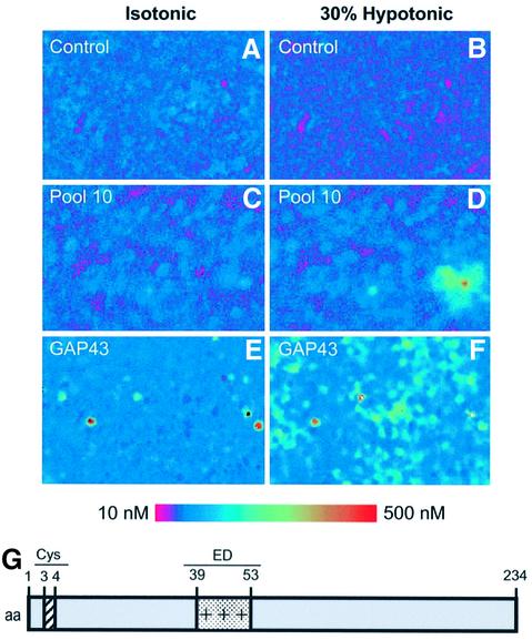
Fig. 1. Expression cloning of GAP43 protein using Ca2+ imaging. HEK293 cells transiently transfected with clones from a human DRG library were subjected to microscopic fluorescent calcium imaging in isotonic (left) and 30% hypotonic conditions (right). Non-transfected cells exhibited no response to the osmotic–mechanical stimulus (A and B). Cells transfected with pool 10 show a marked increase in cytoplasmic calcium (C and D). This pool was subdivided and re-assayed iteratively until a single positive clone (GAP43) was isolated (E and F). Elevated relative Ca2+ concentrations are indicated by an increased ratio of Fura-2 emission at 340 versus 380 nm excitation wavelenght (see calibrated colored bar). (G) Modular organization of GAP43. Palmitoylation occurs at Cys3 and Cys4. The protein displays a positively charged segment (amino acids 39–53) known as the ED domain.

An official website of the United States government
Here's how you know
Official websites use .gov
A
.gov website belongs to an official
government organization in the United States.
Secure .gov websites use HTTPS
A lock (
) or https:// means you've safely
connected to the .gov website. Share sensitive
information only on official, secure websites.
