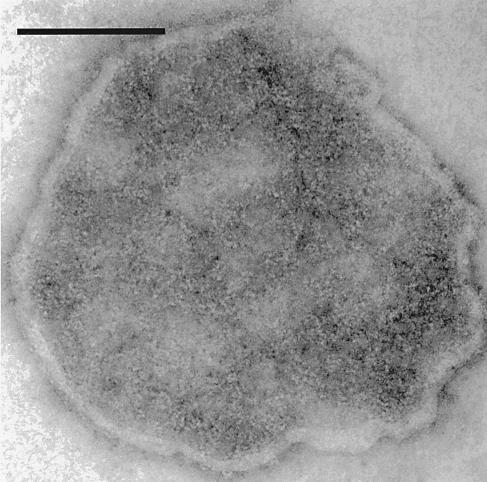
Figure 1.
Electron micrograph of purified Na+,K+-ATPase preparation. A Na+,K+-ATPase preparation suspended in Tris, pH 7.5 buffer was adhered to a formvar-coated copper grid, negatively stained with 1% uranyl acetate and viewed under an 80 KV beam at ×200,000 magnification. Na+,K+-ATPase molecules in this negatively stained preparation appear as white dots. (Scale bar = 0.1 μm.)

