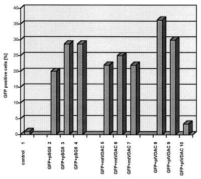
Figure 4.
Cytotoxicity resulting from overexpression of pl-VDAC-1. The percentage of GFP-positive cells after 48 h was quantitated: bar 1, 250 ng of pSG5 expression plasmid without GFP expression cassette; bars 2–4, GFP + pSG5 at ratios of 1:1, 1:3, and 1:5, respectively; bars 5–7, GFP + mt-VDAC-1 at ratios of 1:1, 1:3, and 1:5, respectively; and bars 8–10, GFP + pl-VDAC-1 at ratios of 1:1, 1:3, and 1:5, respectively. A significant dominant-negative effect is evident with a ratio of 1:5 of GFP + pl-VDAC-1 (bar 10), which is not evident after transfection with GFP + mt-VDAC-1 (bar 7).

