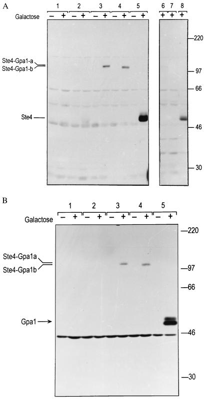
Figure 4.
(a) Western blot showing detection by anti-Ste4 antibody of Ste4-Gpa1 fusions from cells grown in glucose (−) or galactose (+) medium. 40 μg total protein were loaded per lane. Transformants of strain SK1007 (ste4 gpa1), carry the following plasmids: 1, pGT5 (vector); 2, pGT-STE4–1 (STE4); 3, pSTE4-GPA1-a (STE4-GPA1-a); 4, pSTE4-GPA1-b (STE4-GPA1-b); 5, pG1501 (GPA1) and pGT-STE4–2 (STE4). Lanes 6–8 show specific blocking of the anti-Ste4 reactive bands with recombinant Ste4 protein. Lane 6, pSTE4-GPA1-a (STE4-GPA1-a). Lane 7, pSTE4-GPA1-b (STE4-GPA1-b). Lane 8, pG1501 (GPA1) and pGT-STE4–2 (STE4). A number of nonspecific bands are evident, the most prominent migrating at approximately 29, 47, 56, 65, and 110 kDa. The band at 47 kDa seems to be nonspecific, as it appears in the empty vector control, it appears in the absence of galactose, its strength actually decreases in samples grown in galactose, and it is blocked by recombinant Ste4 protein to a much lesser extent (<2-fold) than the bona fide anti-Ste4 reactive products (blocked >10-fold). (b) Western blot by using anti-Gpa1 antibody. Strains as in a. Both myristoylated (lower band) and nonmyristoylated (upper band) forms of Gpa1 are evident. The expected molecular weight of nonmyristoylated Gpa1 is 54 kDa. The band at 45 kDa is nonspecific.

