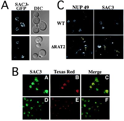
Figure 3.
Sac3-GFP is localized to the nuclear rim and associated with nuclear pore complexes. (A) Localization of Sac3-GFP in living cells (ACY276). (B) Cells (ACY276) expressing Sac3-GFP were prepared for indirect immunofluorescence. Sac3-GFP signal (A and D) is shown in green. Nuclear pores (B) and spindle pole bodies (E) are shown in red. Overlap between the signal for Sac3-GFP and either nuclear pore antigens (C) or spindle pole bodies (F) is indicated by the yellow in the merged images. (C) Wild-type and NUP120/RAT2Δ cells expressing either Sac3-GFP or Nup49-GFP were grown at 25°C and then shifted to the nonpermissive temperature (37°C) for 2 hr. Areas of nuclear pore clustering are indicated by arrows.

