FIG. 4.
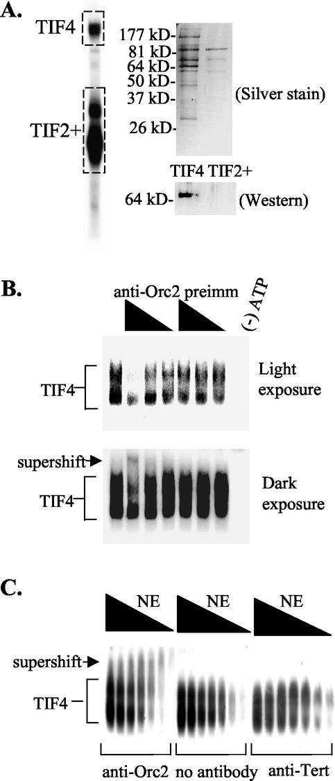
Association of p69 with the TIF4 origin binding complex. (A) Two-dimensional gel analysis of TIF4. EMSA was performed on whole-cell S100 extracts with ssT51. The boxed regions (TIF4 complex and TIF2+ complex) were excised from the EMSA gel. Proteins were electroeluted and separated by SDS-PAGE for silver staining and Western blot analysis with X. laevis Orc2 antibodies. Preimmune serum showed no detectable signal (data not shown; see Fig. 2A), and X. laevis Orc2 antiserum recognized a single 69-kDa polypeptide. (B and C) X. laevis Orc2 antibodies form a ternary complex with TIF4 and the type I element T-rich strand. (B) EMSA analysis with a fixed amount of Q-Sepharose-purified TIF4 and a twofold dilution series of X. laevis Orc2 or preimmune antiserum. Radiolabeled oligonucleotide, ssT51.(C) EMSA analysis with a twofold dilution series of nuclear extracts in the presence and absence of a fixed amount of protein A-Sepharose-purified X. laevis Orc2 or Euplotes crassus telomerase reverse transcriptase (Tert) antibody.

