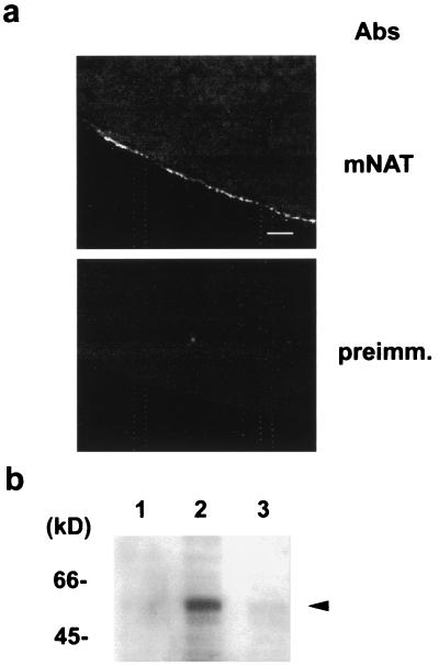
Figure 2.
Expression of mNAT in Xenopus oocytes. (a) Frozen sections of Xenopus laevis oocytes 3 days after injection with cRNA of mNAT were labeled with anti-mNAT antibody or preimmune serum and were examined by confocal microscopy at a scanning interval section of 0.5 μm. (Bar, 30 μm.) (b) Western blot analysis demonstrates overexpression of mNAT (arrrowhead) using a specific antibody in extracts from oocytes injected with water (lane 1), mNAT cRNA (lane 2), or uninjected oocytes (lane 3).

