FIG. 5.
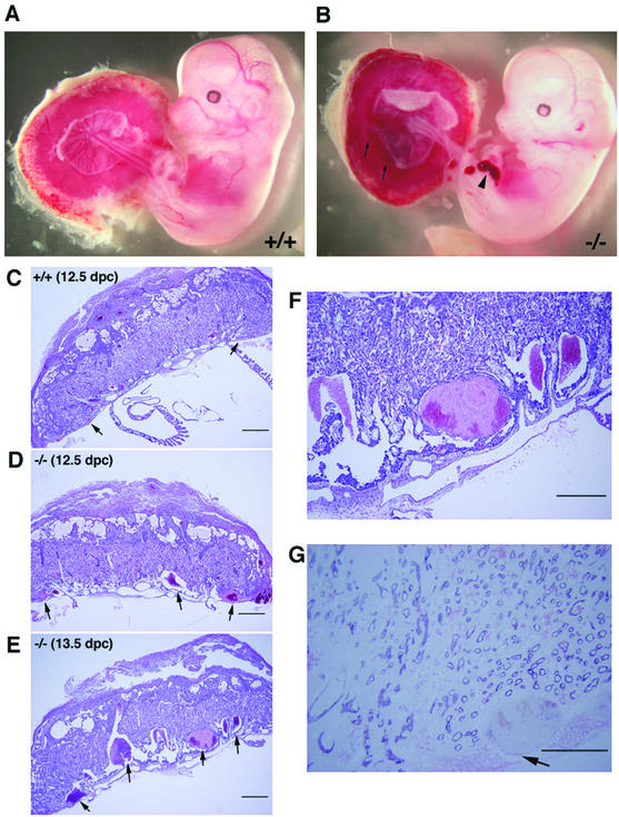
Morphology and histology of wild-type (ROCK-II+/+) and ROCK-II−/− embryos and placentas. (A and B) Embryos isolated together with their placentas. Blood clots found in the ROCK-II−/− placenta (arrows) and hemorrhage in the hind limb of the ROCK-II−/− embryo (arrowhead) are indicated. (C to F) Hematoxylin and eosin staining of a 12.5-dpc wild-type placenta (C), a 12.5-dpc ROCK-II−/− placenta (D), and a 13.5-dpc ROCK-II−/− placenta (E). Panel F shows a higher-magnification view of the labyrinth layer shown in panel E. (G) Immunohistochemical staining of the labyrinth layer of a ROCK-II−/− placenta with an anti-PECAM antibody. The arrow indicates a blood clot found in this section. The absence of nucleated blood cells and PECAM1-positive endothelial cells around the clot indicates that the clot had a maternal origin. Bars, 500 μm (C, D, and E), 300 μm (F), and 200 μm (G).

