Figure 1.
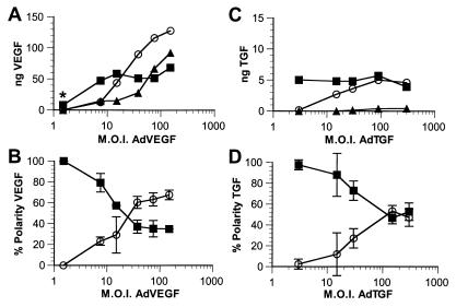
Saturation of apical VEGF and TGF secretion on transduction via adenovirus vectors. RPE-J cells grown on porous filter supports were transduced with AdVEGF (A and B) or AdTGF (C and D) at increasing mois. The amounts of VEGF (A and B) or TGF (C and D) secreted into the apical (■) or basal (○) media, or retained in the cells (▴) were determined by ELISA. A and C show representative experiments demonstrating the amount of VEGF or TGF actually detected in the samples after adjusting for volume. B and D illustrate the percentage of total secreted VEGF or TGF (polarity) released into the apical (■) or the basal (○) media. Note that, although at low mois nearly 100% of both VEGF and TGF are secreted into the apical medium, increasing mois result in large increases in the production of VEGF or TGF with progressively larger fractions secreted basally or retained by the cells. Data in C and D are represented as mean ± SD (n ≥ 3). *, this point is from a separate experiment.

