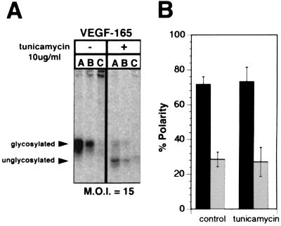
Figure 7.
N-glycans are not necessary for apical secretion of VEGF. The secretion of VEGF by RPE-J cells transduced with AdVEGF at a moi of 15 was examined by pulse–chase analysis as in Fig. 2. Twenty-four hours after transduction with AdVEGF, cells were pretreated for 30 min with 0 or 10 μg/ml tunicamycin (an inhibitor of N-glycosylation), were starved for 30 min in cysteine- and methionine-free medium, and then were labeled with [35S]express and [35S]cysteine for 20 min. After a 3-h chase in medium containing 0 or 10 μg/ml tunicamycin, cells were lysed and VEGF was immunoprecipitated from the apical or basal compartments, or the cells. A shows a representative experiment in which VEGF was immunoprecipitated from the apical medium (A), basal medium (B), or cell lysates (C) and was analyzed by SDS/PAGE and phosphorimaging. B shows the quantification of three experiments, presenting the data as mean ± SD (■, apical medium; □, basal medium). Note that VEGF appears to be delivered predominantly to the apical surface of both treated and untreated control cells. That tunicamycin successfully inhibited the glycosylation of VEGF is demonstrated by the increased mobility of VEGF apparent in A. Quantitation of the apical and basal VEGF is indicated a nearly identical 75% apical polarity of secretion of VEGF by tunicamycin treated and untreated cells.

