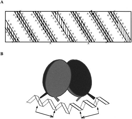
Figure 8.
Model for tetrameric NodD binding on the nod box region. (A) Planar representation of the nodA nod box sequence. The positions of the residues are projected onto the surface of a cylinder that is then unrolled onto a flat surface. The DNA is assumed to adopt the B-form conformation, 10.4 bp per helical turn. The solid arrows indicate the regions protected from DNase I digestion. Closed circles denote the consensus sequence of the D-half (left) and P-half (right). (B) Tetrameric NodD assumes a cyclically symmetric dimer of NodD dimers. Each dimer is also cyclically symmetric and binds to one half-site with the basic motif AT-N10-GAT. Arrows and short lines indicate the exact position of the binding motif T-N11-A-N18-T-N11-A.

