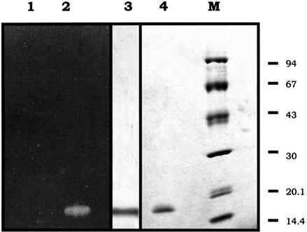
Figure 5.
SDS–PAGE, activity staining and immunoblot analyses of the purified Cc RNase Aliquots of 2 µg of the purified recombinant protein (lane 2) and an equivalent amount of this protein preincubated at 99°C for 5 min (lane 1) were analyzed on an 11–15% polyacrylamide gel containing 0.25 mg/ml poly(C). After electrophoresis the gel was stained for RNase activity as described in Materials and Methods. The same protein amount (lane 3) was analyzed by SDS–PAGE, electro-transferred onto a nitrocellulose membrane and detected using a polyclonal antibody raised against the native poly(U), poly(C)-specific RNase isolated from the insect C.capitata. Purified recombinant Cc RNase (lane 4) and a set of marker proteins of known molecular weight (94, 67, 43, 30, 20.1 and 14.4 kDa top to bottom, lane M) were run in parallel and the proteins were stained with Coomassie blue.

