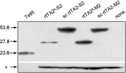
Figure 4.
Western blot of reverse transactivator variants. Ten micrograms of total proteins from HeLa cell extracts were separated by SDS–PAGE. Western blot analysis was performed with anti-TetR polyclonal rabbit serum (laboratory stock) and detected with ECL+ (Amersham). Detection of purified TetR (15 ng) is shown in the first lane. The molecular weights of TetR, rtTA2s-S2, -M2 and those of their sc counterparts are indicated in kDa on the left side. The cellular protein marked with an asterisk was detected with antibody against β-actin and served as an internal loading standard for estimating the amounts of transactivators.

