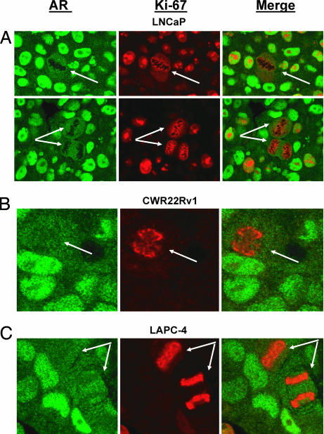
Fig. 5.
AR protein levels are down-regulated during mitosis in AS prostate cancer LNCaP (A), CWR22Rv1 (B), and LAPC-4 (C) xenografts grown in intact male nude mice. In these studies, tumor tissue sections were stained for AR (green) and the proliferation marker Ki-67 (red). Ki-67 is a well established marker for proliferation that is expressed in all parts of cell cycle except in proliferatively quiescent G0 or early G1 phases. Importantly, Ki-67 reacts with an interchromatin network during mitosis, thereby “painting” the chromosomes (36) and making it easy to identify mitotic cells (arrows). Consistent with the in vitro data, AR expression was greatly decreased in cells undergoing mitosis in LNCaP, CWR22Rv1, and LAPC-4 prostate cancer xenografts.

