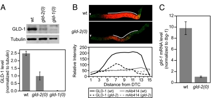
Fig. 3.
GLD-2 regulates gld-1 expression. (A Upper) Western blot of proteins prepared from wild-type, gld-2(0), and gld-1(0) L4 larvae. (Lower) GLD-1 levels in three independent experiments after normalizing to level of tubulin. (B Upper) Germ lines dissected from wild-type and gld-2(0) L4 larvae and stained with anti-GLD-1 (red) and Mab-414 (green), which highlights nuclear pores. Germ lines were treated identically, and confocal images were taken with the same settings at the same magnification. The arrowheads indicate distal ends of germ lines. (Lower) Quantitation of protein abundance in wild type and gld-2(0) mutants. Solid black line, GLD-1 in wild type; dashed black line, GLD-1 in mutant; solid gray line, Mab-414 in wild type; dashed gray line, Mab-414 in mutant. x axis, the distance from the distal tip cell (DTC) in increments of 2 or 3 rows of germ cells. This profile is based on quantitation of the Upper images; similar profiles have been obtained in multiple independent experiments. (C) Real-time PCR analysis of gld-1 mRNA when normalized to tbg-1 mRNA.

