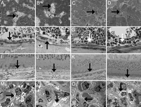
Fig. 3.
Impact of three doses of 1 mg/kg P-GUS on lysosomal storage of P-GUS in liver, bone, retina, and kidney in MR−/− and MR+/+ mice. (A–D) Compared with an untreated MR−/− mouse (A) and an untreated MR+/+ mouse (B), both MR−/− (C) and MR+/+ (D) treated mice had essentially complete clearance of lysosomal storage in the Kupffer cells in the liver (arrow) 1 wk after the last enzyme treatment. (E and F) The bone of untreated MR−/− (E) and MR+/+ (F) mice had lysosomal storage in osteoblasts (arrow) and marrow sinus-lining cells. (G and H) Treated MR−/− (G) and MR+/+ (H) mice had similar marked reduction in storage in the sinus-lining cells. However, only treated MR−/− mice (G) had a marked reduction in storage in the osteoblasts (arrow). Osteoblasts in treated MR+/+ mice (H) had only a slight reduction in storage (arrow). (I and J) Untreated MR−/− (I) and MR+/+ (J) mice had abundant lysosomal storage in retinal pigment epithelial cells (arrow). (K and L) In treated MR−/− mice (K), the retinal pigment epithelial cells had reduced storage. Treated MR+/+ mice (L) showed minimal or no reduction in storage in the retinal pigment epithelial cells. (M and N) Untreated MR−/− (M) and MR+/+ (N) mice both had marked lysosomal distention in the renal visceral epithelial cells (podocytes; arrow). (O and P) After ERT with P-GUS, MR−/− mice (O) showed a marked reduction in lysosomal storage in these cells, but treated MR+/+ mice (P) showed much less treatment effect. (Scale bar: A–L, toluidine blue, 20 μm; M–P, uranyl acetate-lead citrate, 10 μm.)

