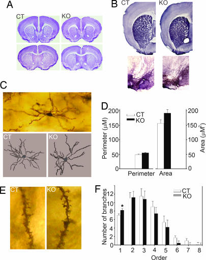
Fig. 2.
Anatomical analysis. (A) Nissl staining showed that KO mice had no gross anatomical abnormalities in the cortex and dorsal or ventral striatum. (B) Dopaminergic innervations to the striatum (Upper) and dopaminergic neurons in the substantia nigra (Lower) also were normal in KO compared with CT mice. (C) A representative medium spiny neuron at ×10 magnification and representative traces of CT and KO medium spiny neurons produced at ×40 magnification. (D) Cell body size of medium spiny neurons of CT and KO mice were similar. (E) No differences in spine count and density were detected between CT and KO mice. Representative second-order dendrites with spines are shown at ×60 magnification. (F) KO mice had significantly more primary dendrites.

