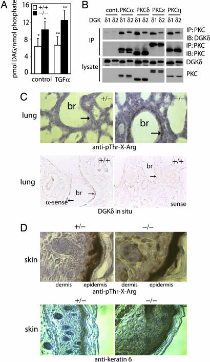
Fig. 5.
DAG levels and assessment of PKC activity. (A) DAG quantity in starved or TGFα-treated (10 ng/ml for 15 min), immortalized embryo fibroblasts was normalized to lipid phosphate. Shown are mean values and SD (n = 4). ∗ and ∗∗ indicate that the changes were significant (P < 0.05) as calculated by one-tailed t tests. (B) FLAG-DGKδ1 or FLAG-DGKδ2 was cotransfected into MCF-7 cells with either control vector or a PKC isotype with a Myc epitope tag. The PKC was immunoprecipitated with anti-Myc antibodies, and coimmunoprecipitation of DGKδ was detected by immunoblotting using anti-FLAG antibodies. (C Upper) Sections from heterozygous (+/−) or DGKδ-null (−/−) newborn mouse lung that were immunostained to detect phospho-Thr-X-Arg and counterstained with hematoxylin. In dgkd−/− mice, there was increased immunostaining (brown, marked by arrows) in the epithelial lining of the major bronchi (br). (C Lower) Lung sections from WT mice processed for in situ hybridization by using an antisense (α-sense) probe. Expression of DGKδ mRNA (brown, marked by arrows) in the epithelium of major bronchi is shown. There was minimal in situ staining of the epithelium (marked by arrow) with the sense probe. (D) Sections from heterozygous (+/−) or DGKδ-null (−/−) newborn mouse back skin immunostained brown to detect phospho-Thr-X-Arg or keratin 6 and counterstained with hematoxylin.

