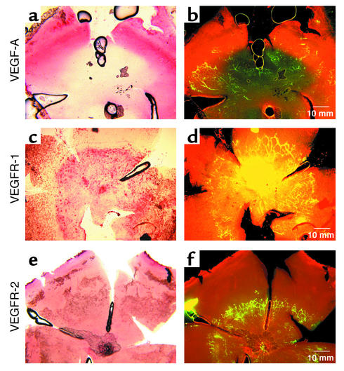
Figure 1.
P5 retinal whole-mount in situ hybridization of VEGF-A, VEGFR-1, and VEGFR-2 mRNA’s. (a) VEGF-A mRNA (pink) is detected anterior to the growing vessel front as outlined by (b) FITC-dextran perfusion of vessels (yellow green). Posterior to the vessel front, VEGF-A expression is suppressed (light yellow). (c) VEGFR-1 mRNA (pink) is detected in the central retina but is not seen at the anterior edge of vessels as outlined by (d) FITC-dextran perfusion of vessels (yellow). (e) VEGFR-2 mRNA (pink) is detected in the entire retina and does not correspond to the vessels (f) outlined in yellow.

