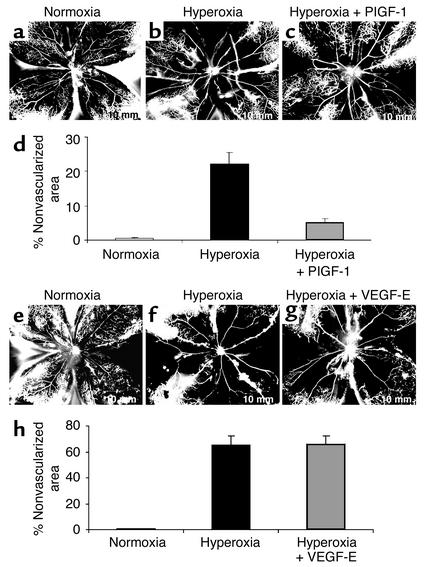
Figure 5.
PlGF-1, but not VEGF-E, prevents hyperoxia-induced retinal vessel loss, thus implicating VEGFR-1 in survival. PlGF-1: P8 FITC-dextran–perfused retinal flat-mount retina from a representative control mouse treated with room air (normoxia) (a) or a mouse given hyperoxic treatment (75% O2 for 17 hours at P7–P8) after intravitreal injection on P7 of (b) control BSS in one eye and (c) the VEGFR-1–specific ligand PlGF-1 in the contralateral eye. Vessels delineated with FITC show that PlGF-1 confers significant protection from oxygen-induced vessel loss compared with BBS control. (d) Analysis of nonvascularized area shows a greater than fourfold difference between eyes treated with PlGF-1 (22.2% ± 3.4% vascularized area) and eyes treated with BSS (5.1% ± 1.2%) (n = 6, P < 0.001). VEGF-E: FITC-dextran–perfused retinal flat mount of P8 control retina from representative room air–treated mouse (e) or oxygen-exposed mouse after intravitreal injections at P7 of (f) control BSS in one eye and (g) VEGFR-2–specific ligand VEGF-E in the contralateral eye. (h) Analysis of nonvascularized area shows no significant difference between VEGF-E– and BSS-treated eyes (n = 6, P = 0.87). Results are representative of two independent experiments.

