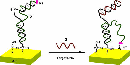
Fig. 1.
Schematic of the signal-on E-DNA sensor, which is based on a conformational change in a MB-modified duplex DNA that occurs after target-induced strand displacement. In the absence of target, the two double-stranded regions formed between the capture and signaling probes sequester the MB from the electrode surface, producing a relatively small MB redox current. When the sensor is challenged with a complementary target, the observed MB redox current increases significantly, presumably because the flexible, single-stranded element liberated in the signaling probe increases the efficiency with which the MB can transfer electrons to the electrode surface. Blue, capture probe (1); green, signaling probe (2); red, target (3).

