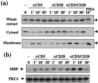
Figure 1.
CD28 costimulation enhances membrane translocation and in situ catalytic activity of PKCθ. (a) Jurkat T cells were stimulated with anti-CD3 and/or anti-CD28 antibodies (2 μg/ml each) for the indicated times. Whole extract, cytosol, and membrane fractions were prepared and resolved by SDS/PAGE, and the expression of PKCθ in each fraction was determined by Western blotting. PMA stimulation (100 ng/ml for 10 min) was used as a positive control for PKC translocation. The position of PKCθ is indicated by arrowheads. (b) Jurkat cells were stimulated as in a. Endogenous PKCθ was immunoprecipitated, and its enzymatic activity was determined (Upper). Kinase reactions were performed in the absence of lipid cofactors or PMA to reflect the in situ activity of PKCθ (11). SDS/PAGE resolved reaction products were analyzed by autoradiography (Upper). The membrane was immunoblotted with a PKCθ-specific antibody (Lower).

