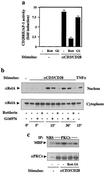
Figure 4.
Inhibition of PKCθ blocks CD28 costimulation. (a) Jurkat T cells were cotransfected with CD28RE/AP-1-Luc (10 μg) and β-galactosidase plasmid (2 μg) reporters. Cells were stimulated for 10 min (20 h later) with CD3- plus CD28-specific antibodies, in the presence or absence of rottlerin (Rott; 30 μM) or Gö6976 (Gö; 0.5 μM). Luciferase activity in cell lysates was determined. (b) Jurkat cells were incubated (15 min at 37°C) in the absence or presence of rottlerin or Gö6976 and then stimulated with anti-CD3/CD28 antibodies or with TNFα (10 ng/ml) for the indicated times. Nuclear and cytoplasmic extracts were prepared, and protein from each fraction (5 μg) was analyzed by Western blotting with an anti-RelA antibody. (c) Wild-type PKCθ-transfected Jurkat T cells were stimulated as in a. Cell lysates were immunoprecipitated (IP) with normal rabbit serum (NRS) or with an anti-PKCθ antibody, and the in vitro enzymatic activity of PKCθ was measured in the presence or absence of rottlerin or Gö6976 (Upper). The membrane was immunoblotted with a PKCθ-specific antibody (Lower).

