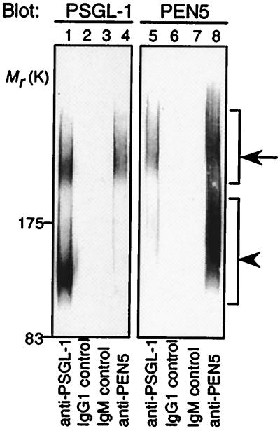
Figure 5.
Biochemical analysis of PSGL-1 and PEN5 on NK cells. NK cell lysates were immunoprecipitated with anti-PSGL-1 mAb (lanes 1 and 5), with control mouse IgG1 (lanes 2 and 6), with control mouse IgM (lanes 3 and 7), or with anti-PEN5 mAb (lanes 4 and 8). Immunoprecipitates were subjected to SDS/PAGE and immunoblotted with anti-PSGL-1 mAb (Left) or anti-PEN5 mAb (Right). The anti-PSGL-1 and anti-PEN5 mAb specifically identified two polydispersed bands of apparent Mr 220–240 kDa (arrow) and 110–140 kDa (arrowhead). Consistent with previous report (31), the relative enhancement of the upper band relative to the lower band is reproducible and is likely the consequence of a higher efficiency of the dimeric PSGL-1 to be included in the immunoprecipitates.

