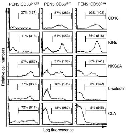
Figure 6.
Coordinated cell surface expression of PEN5 and KIRs on NK cell subsets. Purified NK cells were stained with anti-PEN5, anti-CD56, and anti-L-selectin, anti-CD16, anti-NKG2A, or anti-KIRs mAbs and analyzed by flow cytometry. PEN5−CD56bright, PEN5−CD56dim, and PEN5+CD56dim were analyzed for CD16, KIRs, NKG2A, L-selectin, and CLA expression. Vertical cursor was set with appropriate negative control mAb. The proportions and the mean fluorescence intensity (MFI) of positive cells are indicated in each histograms.

