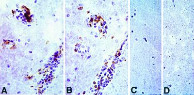
Figure 5.
Infiltration of transgenic Vβ6+ cells in the CNS of a transgenic mouse with spontaneous EAE. A and B are adjacent serial sections immunostained for CD4 (A) and Vβ6 (B) from a 5B6 TCR-transgenic mouse with spontaneous EAE. Similar numbers of perivascular inflammatory cells are stained in each. C and D are sections from the brain of a TCR-transgenic mouse, which did not develop spontaneous EAE, stained for CD4 and Vβ6, respectively. (All sections are stainings with immunoperoxidase with hematoxylin; (×514.)

