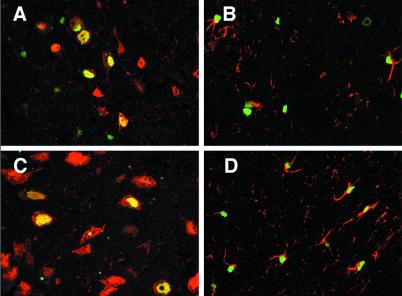
Figure 4.
Identification of transduced cells after intrastriatal injection of rAAV5βgal. Fifteen weeks after injection of rAAV5βgal, coronal brain sections were dual stained for β-galactosidase (green nuclei) and NeuN (neuronal-specific, red nuclei and light red cytoplasm), or for β-galactosidase and GFAP (astrocyte-specific, red cell processes). Confocal microscopy image analysis was performed, and representative two-color-merged images of single z-series slices are shown. In the striatum, both transduced neurons (yellow cell nuclei in A) and transduced astrocytes (B) were detected. In the medial septal region, transduction appeared to be restricted to neurons (C), whereas in the corpus callosum, the transduced cells were GFAP-positive astrocytes (D). Images were captured by using a ×40 (A, B, and D) or ×63 (C) oil-immersion objective.

