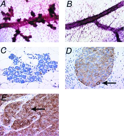
Figure 4.
Digitized micrographic images of whole-mount (A and B) and immunohistochemical (C–E) analysis of the MMTV-Cre Flneo NeuNT mammary gland and tumors. (A) Whole mount of a virgin MMTV-Cre Flneo NeuNT mammary gland at 9 months. The extensive side branching terminating in lobuolalveolar units should be noted. (B) Whole mount of a virgin wild-type control mammary gland at a similar stage of development illustrating a normal duct with few side branches. (C) Immunohistochemical analysis of the same virgin gland shown in A. Note the absence of anti-Neu staining in the acinar structures, which are not dysplastic. Immunohistochemical analysis on the bigenic (D) and a control MMTV-Neu (E) induced tumor shows high levels of Neu expression in these tumors. The contrast in the cytoplasmic and membrane localization of the stain indicated by the arrows in D and E, respectively should be noted. (Magnifications: A and B, × 40; C–E, ×320.)

