
Figure 3.
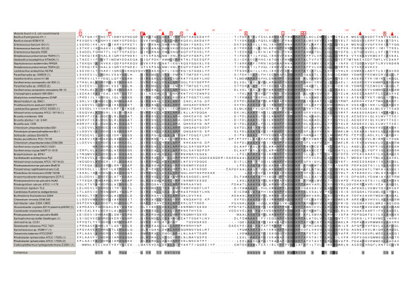
Putative toxins members of the TasAB family members. Multiple alignments of the putative toxins related to the TasB toxin of pGI1 from B. thuringiensis H1.1. Only TasB-like proteins with obvious upstream TasA-like partners (see Fig. 4) were included in this comparison. The left column shows the bacterial host of the protein. Fully conserved amino acids are in dark grey while the other most conserved residues (>50%) are shown in light grey. Variations observed in the TasB mutants recovered from cloning in E. coli are displayed at the top line of the alignment. Many are point mutations (square) and other are early stops (triangle). The consensus sequence is displayed in the last line of the pile-up.