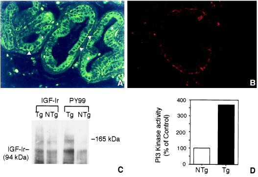
Figure 1.
Indirect immunofluorescence localization of human IGF-1 (A) and mouse IGF-1r (B) in ventral prostate of an 8-week-old BK5.IGF-1 mouse. Arrowheads in A point to the brightly labeled basal epithelial cells, and asterisks are placed on top of luminal cells of the prostatic acini. (A and B, ×300.) (C) Western blot analysis of IGF-1r in prostate of BK5.IGF-1 mice. IGF-1r levels were determined in tissue lysates prepared as described (21) from pooled ventral and dorsolateral prostate of 8- to 10-week-old nontransgenic (NTg) and transgenic (Tg) mice. Protein bands were visualized by enhanced chemiluminescence (ECL, Amersham). (D) Analysis of PI3 kinase activity in prostate tissue (ventral and dorsolateral combined) lysates of 8-week-old nontransgenic (NTg) and transgenic (Tg) mice. The PI3 kinase assay was performed as described (21). In brief, PI3 kinase activity was determined in immunoprecipitates (anti-PI3 kinase antibody, Upstate Biotechnology) by the incorporation of [γ-32P]ATP. Radioactivity was assayed by liquid scintillation counting. Note that nearly identical results were obtained in repeat experiments for both IGF-lr phosphorylation and PI3 kinase activity.

