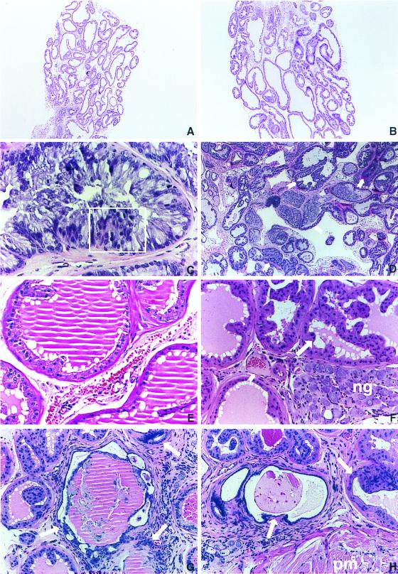
Figure 2.
Preneoplastic and neoplastic changes in the ventral and dorsolateral prostate in BK5.IGF-1 mice. Five-micrometer sections of formalin-fixed tissue embedded in paraffin were stained with hematoxylin and eosin. (A) Ventral prostate of a 7-month-old nontransgenic littermate. (B) A transgenic mouse of the same age. Note that the hyperplasia in the transgenic mouse is sufficient to increase the overall size of the gland in B. (A and B, ×37.5.) (C) Atypical acinus at higher magnification illustrating epithelial stratification (boxed area) as well as both cellular and nuclear atypia. (×300.) (D) Ventral prostate adenocarcinoma from a transgenic mouse illustrating the disorganized growth typical of these lesions. Arrows indicate atypical acini invading periglandular adipose tissue. (×75.) (E) Acini from dorsolateral prostate of a nontransgenic mouse at 14 months of age showing normal appearance. (×150.) (F) Atypical acinus from dorsolateral prostate of a 6-month-old transgenic mouse. Note the presence of medium grade PIN lesions (see long arrow). Neural ganglia (ng) compression caused by gland enlargement and stromal vascularization are also evident in this section. Short arrow points to a highly vascularized area of this lesion. (×150.) (G and H) Focal areas of an adenocarcinoma from a 14-month-old transgenic mouse showing atypical glandular structures and vascularized stroma. Note in H the invasion into the pelvic muscle mass (pm). (G and H, ×150.)

