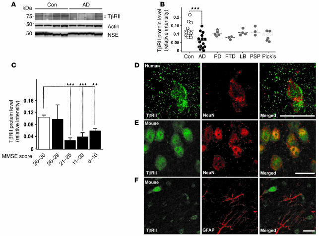
Figure 1. TβRII protein levels are decreased in the human AD brain.
(A) Representative immunoblots of lysates from AD and age-matched control (Con) parietal corteices probed with antibodies against TβRII, actin, and neuron-specific enolase (NSE). (B) TβRII levels of nondemented control, AD, Parkinson’s disease (PD), progressive frontotemporal dementia (FTD), lewy body dementia (LB), progressive supranuclear palsy (PSP), and Pick’s disease (Pick’s) cases were quantified and normalized against protein amounts loaded. Each symbol represents an individual case. (C) TβRII protein levels grouped per mini-mental state exam (MMSE) score (white bar, nondemented controls; black bars, AD). (D and E) TβRII immunofluorescence localized to neurons in the mid-frontal gyrus of the brain from a 69-year-old human (D) and the cortex of a 3-month-old wild-type mouse (E). (F) Little TβRII staining localized to astrocytes in the mouse brain. Scale bars: 10 μm. **P < 0.05, ***P < 0.005; Student’s t test.

