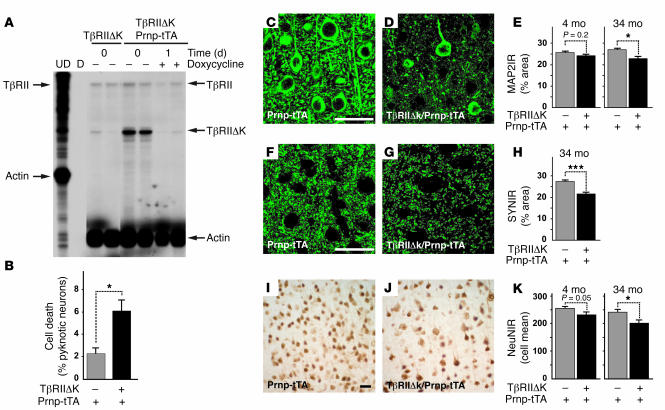
Figure 2. Reduced neuronal TGF-β signaling increases neurodegeneration in vivo.
(A) RNase protection assay shows TβRIIΔk mRNA expression and inhibition of transgene expression in transgenic mice caused by feeding doxycycline-containing food pellets. Arrows on the left indicate undigested (UD) riboprobes; arrows on the right indicate the corresponding protected fragments. TβRII represents the wild-type receptor. D, digested riboprobe. Each lane represents 1 animal. (B) TβRIIΔk-expressing primary neurons show reduced survival in culture. Primary neuron cultures were stained with BODIPY-FL-Fallacidin and Hoechst after 4 days in culture, and the number of viable and dead cells was counted based on the absence or presence of pyknotic nuclei. Data are mean ± SEM of triplicate wells of 3 mice per genotype. (C–K) Sagittal brain sections of 34-month-old (C–K) as well as 4-month-old (E, H, and K) TβRIIΔk/Prnp-tTA or Prnp-tTA mice were immunolabeled for MAP2 (C–E), synaptophysin (F–H), and NeuN (I–K), and percent immunoreactive area (IR) of the neuropil (C–H) or mean number of labeled cells (I–K) in the neocortex was determined by confocal microscopy and computer-aided image analysis. Scale bars: 50 μm. Images are examples from cases with clearly visible degeneration. Data are mean ± SEM (n = 5–7 mice per genotype). *P < 0.05, ***P < 0.005; Student’s t test.

