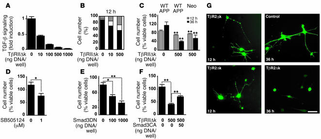
Figure 4. Inhibition of endogenous TGF-β signaling increases neuritic degeneration and Aβ production in neuroblastoma cells.
(A) Transient transfection of TβRIIΔk and a TGF-β reporter plasmid (p800 luciferase) into B103-APPwt cells dose-dependently inhibited signaling 24 hours after transfection. (B) Quantification of the percentage of B103-APPwt cells transiently transfected with TβRIIΔk that showed healthy (black), beaded (white), or no processes (gray) 12 hours after transfection. (C) Expression of TβRIIΔk in B103-APPwt (APPwt) or B103 cells lacking APP (neo) significantly reduced the percentage of cells with healthy processes 12 and 36 hours after transfection. (D–F) Downstream inhibition of TGF-β signaling via treatment with 1 μM SB505124 (D) or expression of dominant-negative Smad3 (Smad3DN; E) reduced the percentage of cells with healthy processes 36 hours after treatment. (F) Expression of constitutively active Smad3 (Smad3CA) partially rescued the phenotype in cells coexpressing TβRIIΔk. (G) B103-APPwt cells expressing TβRIIΔk showed beading and retraction of neurites (12 h), followed by rounding up of the cell body (36 h). Control cells were transfected with filler plasmid and GFP. Scale bar: 50 μm. *P < 0.05, **P < 0.01 versus respective controls; Student’s t test.

