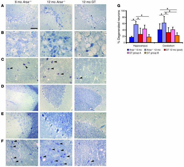
Figure 2. Correction of sulfatide storage and neuronal damage in the CNS of GT-treated mice.
Transverse semithin sections of the hippocampal CA2–3 regions (A and B) and fimbria (C), the cerebellar Purkinje cell layer (D and E), and white matter (F) of untreated and mock-treated Arsa–/– mice (6 and 12 months old, as indicated), and 12-month-old GT mice. Cells with pathological features were already detectable in the pyramidal cell layer of the hippocampus and in the Purkinje cell layer of cerebellum at 6 months (left panels). Lipid storage (arrowheads) was detected throughout the white matter, particularly in the fimbria and cerebellum. The neuronal damage became more severe and the number and size of metachromatic deposits increased significantly in 12-month-old mice (central panels). A marked reduction of sulfatide-containing metachromatic granuli in the white matter and of neuronal damage in CA2–3 and in the Purkinje cell layer was observed in GT mice (right panels). Scale bar: 120 μm (A and D); 80 μm (B, C, and F); and 50 μm (E). (G) Morphometric analysis of neuronal damage, shown as percentage of total counted neurons. Neurons in the CA2–3 and in the Purkinje cell layer were protected from age-related degeneration. In 12-month-old group B treated mice, reduction of degenerating neurons as compared with that of 6-month-old untreated mice was observed, indicating neuronal rescue (*P < 0.05).

