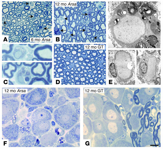
Figure 3. Correction of PNS pathology in GT-treated mice.
Semithin sections and EM images from sciatic nerve and DRG are shown. (A) Metachromatic deposits (arrows) were detected in the SC cytoplasm of 6-month-old untreated Arsa–/– mice. (B) By 12 months of age, the number and size of metachromatic deposits in SCs increased, and demyelination of nerve fibers became more apparent (arrowhead), as also shown at higher magnification in C and in detail by EM in E. (D) In GT mice, neither sulfatide storage nor demyelination was observed. (F) Metachromatic deposits and intracytoplasmatic vacuolation were present in sensory neurons as well as in satellite cells and SCs in DRG of 12-month-old Arsa–/– mice. (G) In GT mice, these alterations were almost completely absent. Scale bar: 20 μm (A, B, D, F, and G); 5 μm (C); and 1.5 μm (E).

